Retinal Surgery
The retina is a thin layer of tissue that lines the back of the eye. Images focused on the retina are converted to neural signals that are transmitted to the brain by the optic nerve.
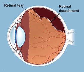
The clear vitreous gel that fills the back of the eye becomes more liquid as we age and eventually separated from the retina. This separation can cause a retinal hole or tear leading to a retinal detachment (where the retinal tissue separates from the back of the eye).

Retinal detachments are surgically repaired by a retinal specialist. If the detachment is small and the hole is superior, a gas bubble can be place in the eye to push the retina back into position and laser is applied to create scars that will hold the retina in place. This is called pneumatic retinopexy. Larger retinal detachments require a surgical procedure in the operating room. Often, the vitreous gel is removed (vitrectomy) and a band (scleral buckle) is placed around the eye to treat the detachment.
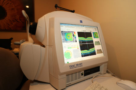
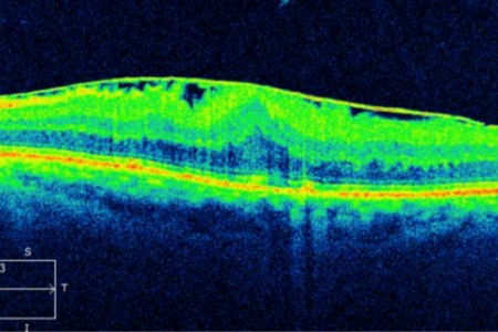
We use Macular Optical Coherence Technology (MOCT) to diagnose and monitor many retinal diseases. This device provides a very detailed image of the retina.

Macular pucker (also caller epiretinal membrane or surface wrinkling retinopathy) is a process where the retina is pulled into folds by a membrane, or scar tissue, on its surface causing distortion of the normally smooth retinal structure. If the retina is thickened and distorted, the images sent to the brain are blurred and distorted. Retinal surgeons treat Macular Pucker by first removing the vitreous gel to have access to the retinal surface. Then, the thin layer of epiretinal membrane (scar tissue) on the surface of the retina is remove and the retina will be smooth and flat again.

A macular hole is a small hole in the center of the retina, right in the center of your vision. These holes can be repaired surgically. The retina surgeon removes the vitreous and places a gas bubble in the eye. The gas is slowly reabsorbed over a few weeks and the hole is most often repaired, restoring vision.
These are just a few examples of retinal problems that are repaired surgically. Retina surgery is only available locally at our surgery center, The Physicians Outpatient Surgery Center, Ltd in Belpre, Ohio.
